Our work “Super-resolved Imaging of a Single Cold Atom on a Nanosecond Timescale” has been published in Physical Review Letters. We demonstrate a groundbreaking method to image a single trapped ion with unprecedented spatial and temporal resolution, breaking through the optical diffraction limit.
Breaking the Diffraction Limit
High-resolution optical detection and imaging of individual particles in cold atomic systems is essential for directly probing quantum phenomena such as transport, correlations, and phase transitions. However, traditional microscopy techniques are fundamentally restricted by the wavelength-scale diffraction limit, making them unsuitable for studying wave function details of many-body systems. We brought the concept of super-resolved microscopy — which has matured as a powerful tool in biology and chemistry — into the realm of cold atoms.
Our Method: Ground-State Depletion Microscopy
We demonstrated ground-state depletion (GSD) microscopy on a trapped-ion system for the first time. By combining the GSD control sequence with the qubit-state polarization and detection methods of 171Yb+ ions, we achieved 2D super-resolved imaging beyond the optical diffraction limit. The key innovation is using a high-purity doughnut-shaped laser beam, generated via a digital micromirror device (DMD) with in-situ aberration correction, to selectively deplete the ion’s quantum state.
Results
We achieved a spatial resolution of 175 nm using a NA = 0.1 imaging objective — a 13-fold improvement compared with direct fluorescence imaging. Moreover, the depletion process can be as short as ~50 ns under strong laser power, enabling us to take stroboscopic images on nanosecond timescales. We demonstrated this capability by observing one complete cycle of the ion’s secular motion within 735 ns, achieving a displacement detection sensitivity of 10 nm.
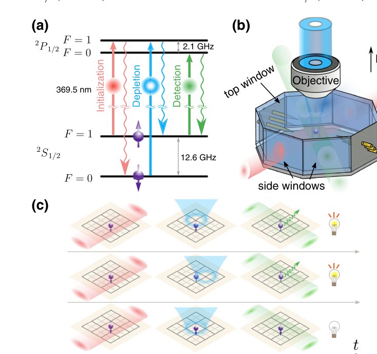
Fig. 1. Experimental scheme and setup. (a) Energy levels of 171Yb+ and the three-laser GSD imaging sequence. (b) A Paul trap with the doughnut beam focused through the top window. (c) Schematic of the super-resolved imaging process: scanning the doughnut spot pixel by pixel to build a super-resolved image.
Our method provides a powerful tool for probing particle positions, momenta, correlations, and dynamics in cold atomic systems, and can be readily extended to neutral atoms in optical tweezers.
Find the article here: https://doi.org/10.1103/PhysRevLett.127.263603