USTC IonQNet Group logo USTCIonQNetGroup
  • Home
  • Research introduction
    Trapped-ion quantum computing node Quantum network with cavity QED Hybrid quantum network
  • Members
  • Publications
  • News
  • Address Join Us Github
  • Categories
    • English
    • 中文

Quantum interface

Cavity QED Interface
From gold-coated fiber mirrors to needle-trap integration: building high-bandwidth, high-efficiency ion–photon interfaces.
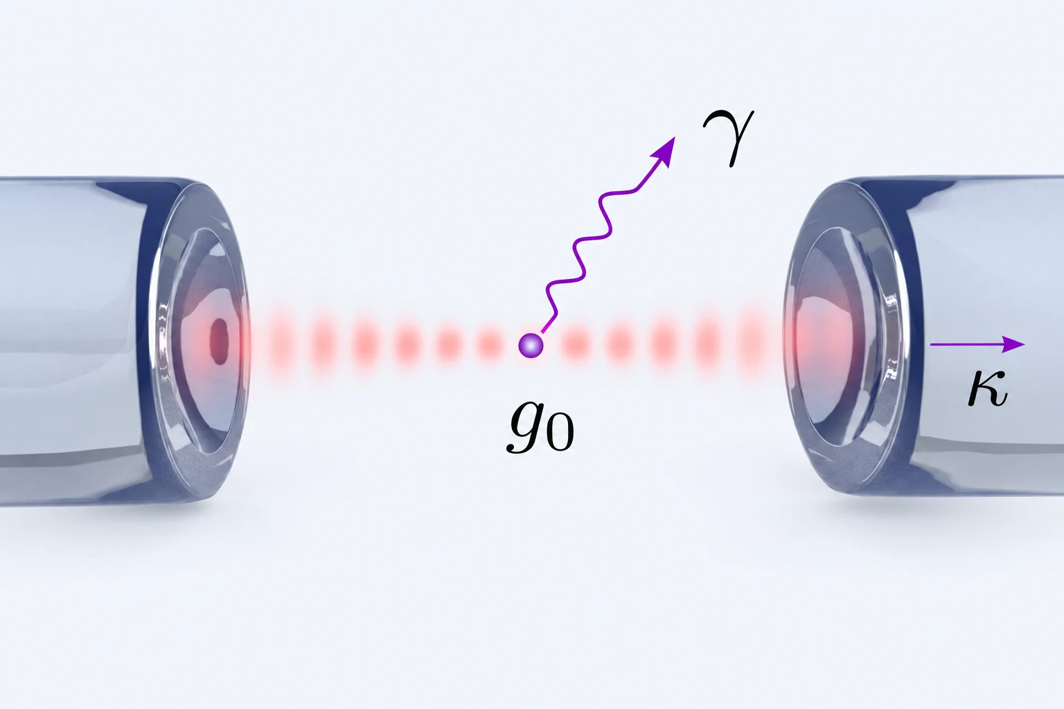
An ion coupled to a fiber microcavity, showing ion–cavity coupling and the emission channels.

We integrate fiber microcavities with needle ion traps to develop ion–photon interfaces for quantum networks. The small cavity mode volume enhances ion–cavity coupling, while the fibers provide channels for collecting and transmitting photons. By designing the mirrors, trapping geometry and optical coupling together, we aim to build network nodes that combine high bandwidth with high efficiency.

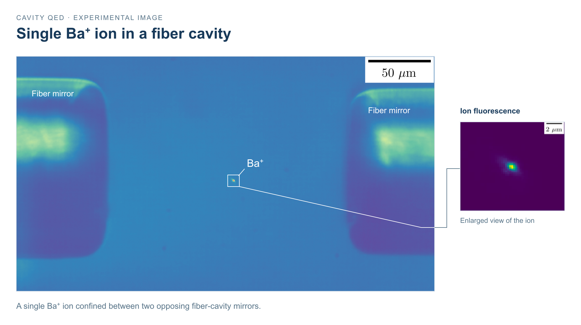
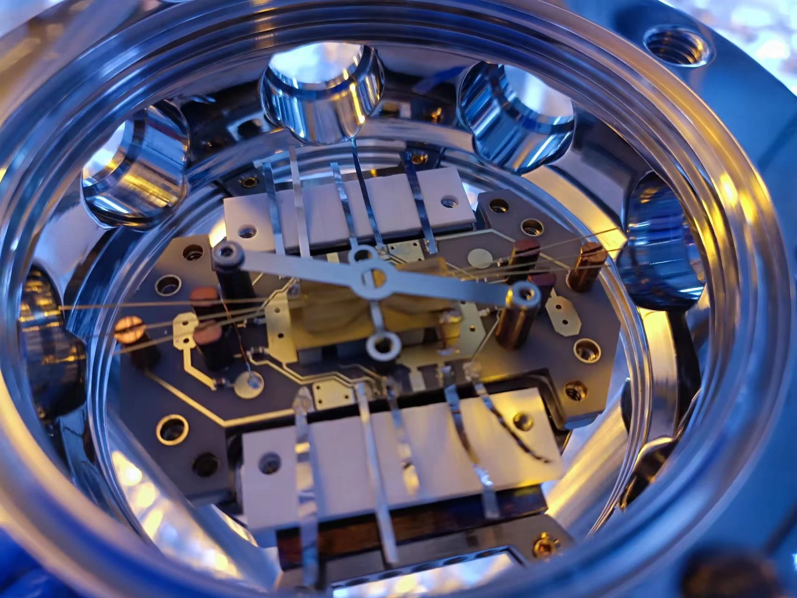
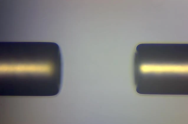
The photographs show the hardware behind this interface. Gold coating on the fiber sides and the outer regions of the end faces provides electrical shielding while leaving the central optical apertures open. Two opposing fiber mirrors form a microcavity, which is integrated with the needle trap inside the vacuum chamber. The fluorescence image of a single Ba⁺ ion between the mirrors shows the ion confined within this cavity geometry.
Explore quantum networking with cavity QED →

Assembly of a fiber cavity integrated with a needle ion trap inside the vacuum chamber.
Two gold-coated fiber-cavity mirrors assembled face to face to form an optical microcavity.
A single Ba⁺ ion trapped between two fiber-cavity mirrors, with an enlarged fluorescence view at right. Scale bars are 50 μm in the overview and 2 μm in the close-up.

Image viewer

  • Back to top
  • USTC
  • LQCC
  • GitHub
  • Atom
  • RSS

Made by Jin-Ming Cui.